38 onion cells under microscope with labels
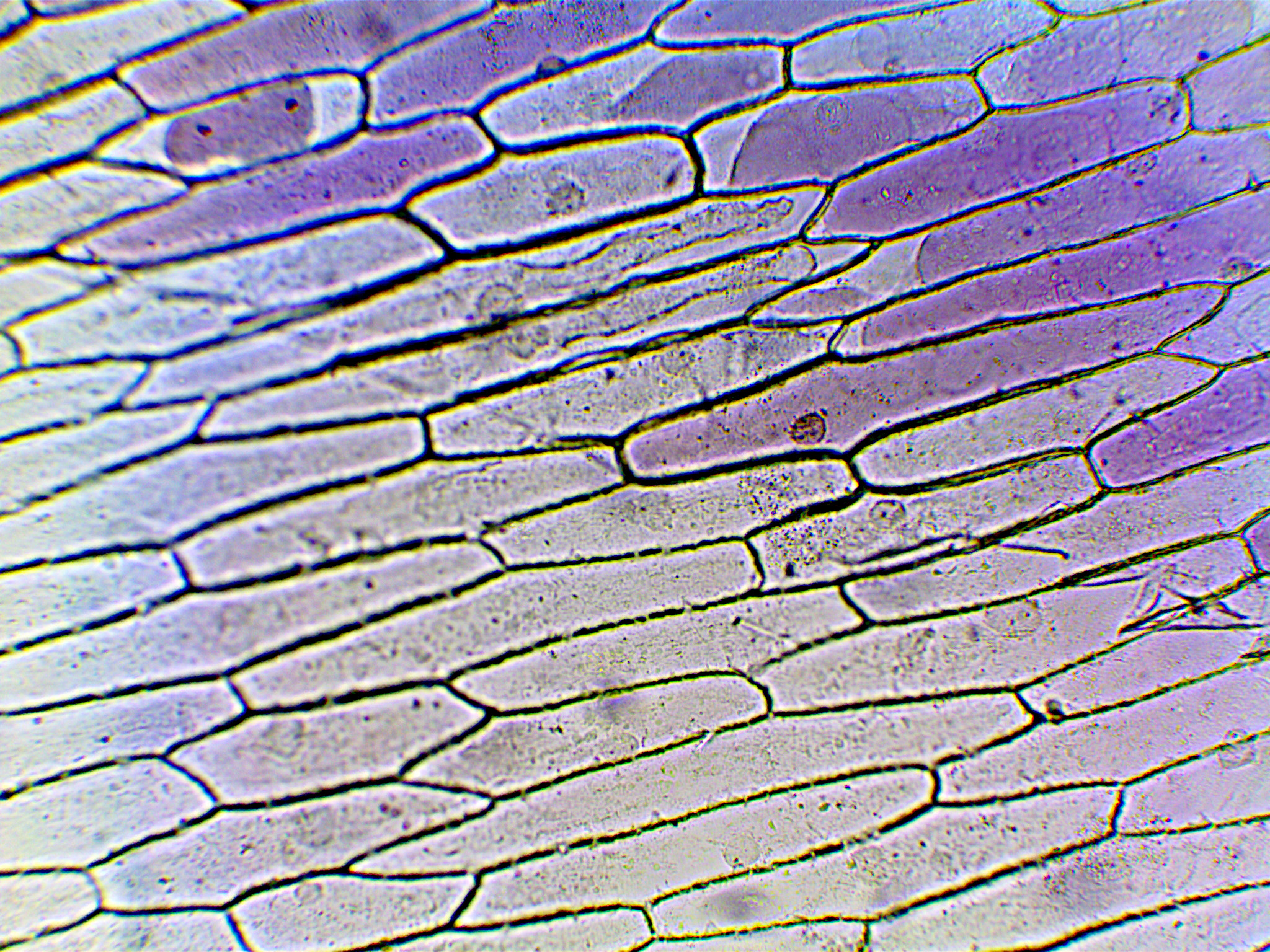
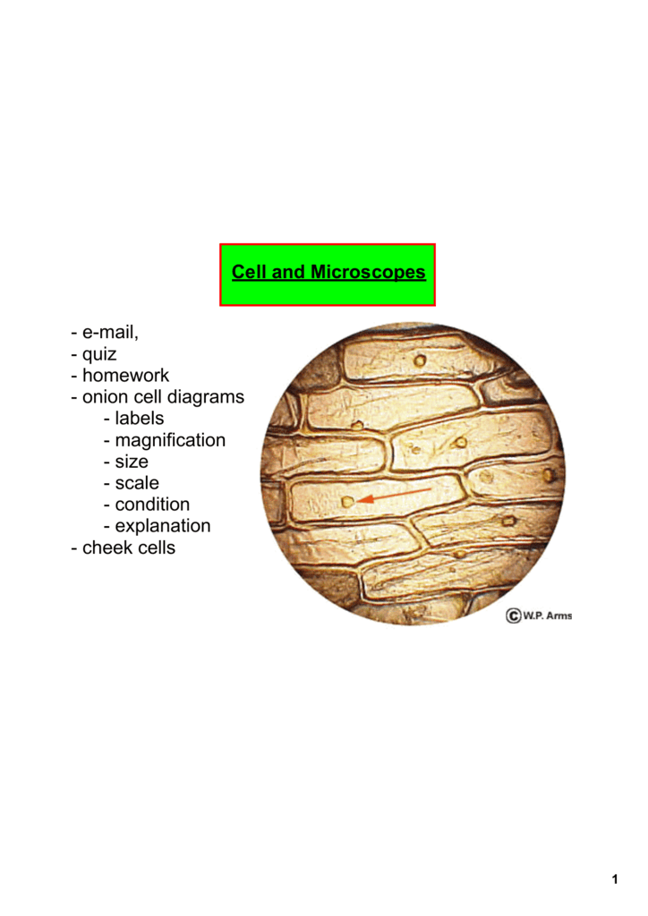
onion cells under a microscope labeled - cahierdeseoul.com RM BRC1AJ - Light Micrograph (LM) of onion skin cells, magnification x 600. Pay close attention, you'll need to label cell slides on the test. Clean the stain from the slide and cover glass. Observe the onion tissue under the microscope at 4x, 10x and 40x with lots of light (open diaphragm). 7. 2. Onion cells under the microscope. Microscope Cell Lab: Cheek, Onion, Zebrina - SchoolWorkHelper The first lab exercise was observing animal cells, in this case, my cheek cells. The second lab exercise was observing plant cells, in this case, onion epidermis. The third lab exercise was observing chloroplasts and biological crystals, in this case, a thin section from the Zebrina plant. The first thing that was done in this lab exercise was ...
vii Sketch the onion peel cell as seen under the microscope Label the ... Put a drop of water in its centre and transfer the peel from the petridish to the slide with the help of a brush. Place the coverslip. (viii) Remove the extra water by placing the slide within a folded filter paper. (ix) Examine the slide first under low power and then under high power. (x) Record your observations.

Onion cells under microscope with labels
Plant Cell Under Microscope Labeled 40X : Young Root 2 Of Broad Bean ... Cells and viewing them under the microscope. A small square of a red onion skin (membrane) was observed under a microscope at high power (x40) magnification. (iv) describe how you applied the stain. They must draw and label the nucleus, cell membrane set up your microscope, place the onion root slide on the stage and focus on low (40x) power. Under the Micrsocope: Onion Cell (100x - 400x) - YouTube In this "experiment" we will see onion cells under the microscope.For the experiment you will only need onion, dropper and the microscope (container and tool... Biology Project The Biology Project, an interactive online resource for learning biology developed at The University of Arizona. The Biology Project is fun, richly illustrated, and tested on 1000s of students.
Onion cells under microscope with labels. Onion Microscope Under Cell Labeled Search: Onion Cell Under Microscope Labeled. Add 2 drops of iodine (or other stain) to the onion slide Use the microscope, slide of "JF", mm ruler and photo of onion cells to assist you in answering the questions You will first view the cell under normal conditions, so you can easily be compared to the results if a change occurs Sketch of one cell in each phase of mitosis (prophase, metaphase ... › Health_Safety_Meeting_DatesHealth & Safety Meeting Dates | Institute Of Infectious ... Feb 08, 2022 · IDM H&S committee meetings for 2022 will be held via Microsoft Teams on the following Tuesdays at 12h30-13h30: 8 February 2022; 31 May 2022; 2 August 2022 Cells and Reproduction - BBC Bitesize All living organisms are made up of cells. Cells are the building units of life - the basic building blocks of all animals and plants. They are so small, you need to use a light microscope to see ... The Biology Project The Biology Project, an interactive online resource for learning biology developed at The University of Arizona. The Biology Project is fun, richly illustrated, and tested on 1000s of students. It has been designed for biology students at the college and high school level, but is useful for medical students, physicians, science writers, and all types of interested people.
Microscopy, size and magnification - Microscopy, size and ... - BBC Place cells on a microscope slide. Add a drop of water or iodine (a chemical stain). Lower a coverslip onto the onion cells using forceps or a mounted needle. This needs to be done gently to... Educational 01: To use a light microscope; 02: To obtain a good specimen of plant tissue for viewing under the microscope (onion cells) 03: To obtain a good specimen of animal tissue for viewing under the microscope (cheek cells) 04: To investigate the digestion of starch by amylase; 05: To investigate the effect of exercise on heart rate ocr.org.uk › Images › 643844-question-paper-depth-inOxford Cambridge and RSA Friday 16 October 2020 – Morning 1 (a) A student was observing onion epithelial cells using a light microscope. They photographed these cells and the image obtained is shown in Fig. 1.1. The student then made a drawing of a few cells from this image. The drawing is shown in Fig. 1.2. Fig. 1.1 cytoplasm cell wall large permanent vacuole ribosome Fig. 1.2 Health & Safety Meeting Dates | Institute Of Infectious Disease … 08.02.2022 · IDM H&S committee meetings for 2022 will be held via Microsoft Teams on the following Tuesdays at 12h30-13h30: 8 February 2022; 31 May 2022; 2 August 2022
ONION CELLS VIDEO - YouTube Video shows how to make a wet mount slide to view onion cells under the microscope. Lennox Educational 01: To use a light microscope; 02: To obtain a good specimen of plant tissue for viewing under the microscope (onion cells) 03: To obtain a good specimen of animal tissue for viewing under the microscope (cheek cells) 04: To investigate the digestion of starch by amylase; 05: To investigate the effect of exercise on heart rate PDF Onion Cell Lab - somewaresinmaine.com Research Biology Onion Cell Lab page 1 of 3 Onion Cell Lab After you have completed the rest of this lab come back to this cover page DRAW & LABEL AN ONION CELL WITH ALL THE PARTS / ORGANELLES YOU OBSERVE UNDER 40X. Purpose: To observe and identify major plant cell structures and to relate the structure of the cell to its function. Materials: 1 ... OBSERVING ONION PEEL EPIDERMAL CELLS UNDER MICROSCOPE - YouTube This video is specially dedicated for my hindi subscribers. Mr Devbrat is one of them. Thank You for watching this video.This video demonstrates how to see e...
What organelles are in an onion cell? - Biology Stack Exchange You cannot see most of these as they appear translucent as well as being too small to see under the light microscope. You need an electron microscope to view these. Note: chloroplasts are not present in an onion cell as it is not a photosynthesising cell. This is a typical onion cell slide with labels:

Onion Cell Under Microscope - Personal Experience with Microscopes - AyushiSinhaMicroscopy ...
Onion Cells Under a Microscope - Requirements/Preparation/Observation Add a drop of iodine solution on the onion membrane (or methylene blue) Gently lay a microscopic cover slip on the membrane and press it down gently using a needle to remove air bubbles. Touch a blotting paper on one side of the slide to drain excess iodine/water solution, Place the slide on the microscope stage under low power to observe.
The Cell - ScienceQuiz.net The diagram shows a group of onion cells. The parts labelled A, B and C respectively are? A = cell wall, B = cytoplasm, C = nucleus ? A = cell wall, B = cytoplasm, C = membrane? A = cytoplasm, B = cell wall, C = nucleus? A = nucleus, B = cell wall, C = cytoplasm; Which one of the following is NOT an example of animal tissue?? Vitamins? Skin? Muscle? Bone; Which one …
Microscope Labeled Cell Under Leaf Endothelial cells under the microscope Images show adaxial epidermis, palisade mesophyll and abaxial Label the cell wall, cytoplasm and nucleus as they appear under high power on page 38 of your manual Procedure: Letter "e" 1 Images were taken on an inverted compound microscope using a 40x DIC objective and digital camera Images were taken ...
Natural Sciences Grade 9 - Grade 7-9 Workbooks Robert Hooke (1635 - 1703). Robert Hooke was the first cytologist to identify cells under his microscope in 1665. He decided to call the microscopic shapes that he saw in a slice of cork "cells" because the shapes reminded him of the cells (rooms) that the monks in the nearby monastery lived in.Robert Hooke was the first to use the term 'cell' when he studied thin slices …
The following diagram shows cells of onion peel label class ... - Vedantu 115.2k + views. Hint: The diagrams mentioned above are the internal structure of an onion peel and human cheek cells. In order to label them, we need to understand its anatomy and know about various structures present in it. Onion peel is an example of a plant cell whereas a human cheek cell is an example of an animal cell. Complete answer:
Onion Cell Lab Report.docx - Onion Cell Lab Report By station, remove the single layer of epidermal cells from inner side of the scale leaf. 3(Place the single layer of onion on a glass slide. 4(Place a drop of iodine stain on your onion tissue. 5(Put the cover slip on the stained tissue and gently tap out any air bubbles. 6(Observe the cells under the microscope and see you results.
Cambridge International AS and A Level Biology Coursebook … Enter the email address you signed up with and we'll email you a reset link.
Onion cell Images, Stock Photos & Vectors - Shutterstock Find Onion cell stock images in HD and millions of other royalty-free stock photos, illustrations and vectors in the Shutterstock collection. Thousands of new, high-quality pictures added every day.

Phases of mitosis in control and 1-day CA-treated root tip cells. Bars... | Download Scientific ...
› natural-sciences › gr9Natural Sciences Grade 9 - Grade 7-9 Workbooks The onion cells have a thick cell wall and a cell membrane. The animal cells only have a cell membrane. The onion cells have a regular shape whereas the cheek cells have a irregular shape and seem more flimsy. In the onion cells they might notice a large vacuole which might not be as visible in the cheek cells. Cheek cells do not have vacuoles ...
interxnet.it Click and drag the label to the corresponding cell under the microscope. 5) 53 (52) 3-4 17 (41. The objective of this experiment was to calculate the percentage of cells in each of the phases of mitosis. The cell cycle checkpoint proteins were analysed with respect to detailed clinicopathological information available for all patients in this cohort. 3. When the …
› bitesize › articlesCells and Reproduction - BBC Bitesize Onion cells are easy to see using a light microscope. ... A small tube placed under the skin of the upper arm. ... Five small tubes with labels and stoppers or lids Cress seeds Labels Cotton wool ...
Onion Cells Under a Microscope (100x-2500x) - YouTube In this video you will see onion cells under a microscope (100x-2500x) as is, without any coloring. To observe the onion cells the thin membrane is used. It...
VIEWING PLANT CELLS UNDER THE MICROSCOPE: onion ... MICROSCOPE: onion cell preparation. This method allows students to view plant cells under the microscope. A single layer of onion.2 pages


Post a Comment for "38 onion cells under microscope with labels"