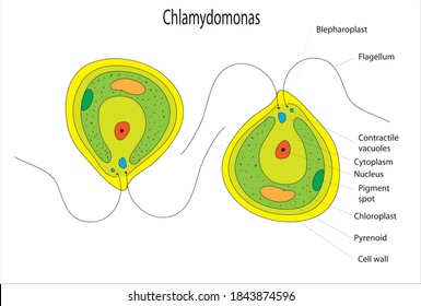
39 chlamydomonas diagram with labels
› lifestyleLifestyle | Daily Life | News | The Sydney Morning Herald The latest Lifestyle | Daily Life news, tips, opinion and advice from The Sydney Morning Herald covering life and relationships, beauty, fashion, health & wellbeing Biological drawings. Structure of Chlamydomonas. Learning ... Structure of Chlamydomonas: Next Drawing > Chlamydomonas is the name given to a genus of microscopic, unicellular green plants (algae) which live in fresh water. Typically their single-cell body is approximately spherical, about 0.02 mm across, with a cell wall surrounding the cytoplasm and a central nucleus.
Chlamydomonas | Facts, Structure, Life Cycle ... Chlamydomonas, genus of biflagellated single-celled green algae (family Chlamydomonadaceae) found in soil, ponds, and ditches. Chlamydomonas species can become so abundant as to colour fresh water green, and one species, C. nivalis, contains a red pigment known as hematochrome, which sometimes imparts a red colour to melting snow. red snow
Chlamydomonas diagram with labels
Asymmetric properties of the Chlamydomonas reinhardtii ... The C. reinhardtii eyespot. (a) A diagram illustrating asymmetric localization of the eyespot relative to the cytoskeleton. Two flagella and four microtubule rootlets extend from a pair of basal bodies at the anterior end of the cell; both the mother basal body (small black oval) and the daughter basal body (small gray oval) are associated with a four-membered rootlet (M4 or D4) and a two ... How to make label Diagram of chlamydomonas - YouTube watch: "How to make thumbnail our you tube videos Hindi /urdu haris by #Top2utv" ... LABORATORY 9 - comenius.susqu.edu Labeled diagram of Chlamydomonas. ... Chlamydomonas from culture. Cells have been stained with Lugol's Iodine, which complexes with true starch to turn black. 400X . You have slides of colonial volvocine green algae, which include Volvox, Gonium , Eudorina, ...
Chlamydomonas diagram with labels. Eye Diagram With Labels and detailed description A brief description of the eye along with a well-labelled diagram is given below for reference. Well-Labelled Diagram of Eye The anterior chamber of the eye is the space between the cornea and the iris and is filled with a lubricating fluid, aqueous humour. The vascular layer of the eye, known as the choroid contains the connective tissue. Diagram the life cycles of Chlamydomonas, Ulothrix ... Solution for Diagram the life cycles of Chlamydomonas, Ulothrix, Spirogyra, and Oedogonium; indicate where meiosis and fertilization occur in each. close. Start your trial now! First week only $4.99! arrow_forward. learn. write. tutor. study resourcesexpand_more. Study Resources. We've got the study and writing resources you need for your ... Life Cycle of Chlamydomonas (With Diagram) ADVERTISEMENTS: In this article we will discuss about the asexual and sexual methods of reproduction that occur in the life cycle of chlamydomonas. 1. Asexual Reproduction: It takes place by following methods: (A) By zoospores- The zoospore formation takes place during favourable conditions. The zoospore formation takes place as follows: ADVERTISEMENTS: The protoplast contracts and […] Chloroplast Structure and Function in detail with Labelled ... Chloroplast Structure and Function in detail with Labelled Diagram. The leaves of the trees are colored in different shades of green. This color is imparted due to the presence of various pigments present in the Chloroplast leaves. The chloroplasts are the cell organelles which consist of these pigments.
› de › financeFinances in Germany - Expat Guide to Germany | Expatica Understanding your money management options as an expat living in Germany can be tricky. From opening a bank account to insuring your family’s home and belongings, it’s important you know which options are right for you. Solved: Label this diagram of the Chlamydomonas life cycle ... LearnSmart Online for Biology (10th Edition) Edit edition. This problem has been solved: Solutions for Chapter 21 Problem 24TY: Label this. diagram of the. Chlamydomonas life cycle.…. Get solutions. Get solutions Get solutions done loading. Looking for the textbook? catalog.jccc.edu › coursedescriptions › biolBiology (BIOL) < Johnson County Community College BIOL 135 Principles of Cell and Molecular Biology (4 Hours). This course is for biology majors and students planning to take additional courses in the life sciences. Subjects covered include the nature of science; the levels of organization and emergent properties of life; basic biochemistry and bioenergetics; cell structure and function; cellular reproduction; Mendelian and molecular genetics ... Chlamydomonas: Position, Occurrence and Structure (With ... Chlamydomonas is unicellular, motile green algae. The thallus is represented by a single cell. It is about 20 p,-30|i in length and 20 µ in diameter. The shape of thallus can be oval, spherical, oblong, ellipsoidal or pyriform. The pyriform or pear shaped thalli are common, they have narrow anterior end and a broad posterior end (Fig. 1).
Structure of Chlamydomonas (With Diagram) | Genetics In this article we will discuss about the structure of chlamydomonas (explained with labelled diagram). The unicellular green alga Chlamydomonas is haploid with a single nucleus, a chloroplast and several mitochondria (Fig. 9.3). It can reproduce asexually as well as sexually by fusion of gametes of opposite mating types (mt + and mt - ). Structure of Chlamydomonas (With Diagram) | Chlorophyta In this article we will discuss about the structure of chlamydomonas with the help of suitable diagrams. Chlamydomonas is unicellular, motile green algae. The thallus is represented by a single cell. It is about 20 p,-30|i in length and 20 µ in diameter. The shape of thallus can be oval, spherical, oblong, ellipsoidal or pyriform. Diagram of Chlamydomonas angulosa, Flagellated Protozoan ... Diagram of Chlamydomonas angulosa, Flagellated Protozoan. Drawing. Get premium, high resolution news photos at Getty Images Describe the structure of chlamydomonas with neat labelled ... answeredOct 30, 2020by Naaji(56.8kpoints) selectedOct 30, 2020by Jaini Best answer 1. Chlamydomonas is a simple, unicellular, motile fresh water algae. They are oval, spherical or pyriform in shape. 2. The cell is surrounded by a thin and firm cell wall made of cellulose. 3. The cytoplasm is seen in between the cell membrane and the chloroplast. 4.
Labeled Paramecium Diagram by ScienceDoodles on DeviantArt Paramecium are the coolest. Simply rad. Unbeatable in every way shape or form.
› pmc › articlesActin and Actin-Binding Proteins - PMC A seed was first decorated with myosin heads and then allowed to grow bare extensions. Elongation was faster at the barbed end than at the pointed end. (B) Diagram showing the rate constants for actin association and dissociation at the two ends of an actin filament. The pointed end is at the top and the barbed end is at the bottom.
Draw a neat labelled diagram. Chlamydomonas - Biology ... Draw a neat labelled diagram. Chlamydomonas . Maharashtra State Board HSC Science (General) 11th. Textbook Solutions 8018. Important Solutions 19. Question Bank Solutions 5545. Concept Notes & Videos 420. Syllabus. Advertisement Remove all ads. Draw a neat labelled diagram. ...
Animal Cells: Labelled Diagram, Definitions, and Structure Animal Cells Organelles and Functions. A double layer that supports and protects the cell. Allows materials in and out. The control center of the cell. Nucleus contains majority of cell's the DNA. Popularly known as the "Powerhouse". Breaks down food to produce energy in the form of ATP.
Morphology of Chlamydomonas (With Diagram) | Algae In this article we will discuss about the external morphology of chlamydomonas. Also learn about its Neuromotor Apparatus, Electron Micrograph, Palmella-Stage with suitable diagram. 1. The organism is an unicellular alga (Fig. 11). 2. The thallus is spherical to oblong in shape but some species are pyriform or ovoid. ADVERTISEMENTS: 3.
Chlamydomonas reinhardtii - microbewiki Chlamydomonas reinhardtii features are ovate in shape, about 10 um, unicellular with a distinct cell wall, and a single chloroplast in close proximity to the nucleus. The nucleus is typically located in the center and with a distinct nucleolus. There is an eyespot and one or several contractile vacuoles.
Chegg.com | Biology plants, Biology lessons, Science biology May 12, 2015 - Answer to Label this diagram of the Chlamydomonas life cycle.. May 12, 2015 - Answer to Label this diagram of the Chlamydomonas life cycle.. Pinterest. Today. Explore. When autocomplete results are available use up and down arrows to review and enter to select. Touch device users, explore by touch or with swipe gestures.
Chlamydomonas - Wikipedia Chlamydomonas is a genus of green algae consisting of about 150 species all unicellular flagellates, found in stagnant water and on damp soil, in freshwater, seawater, and even in snow as "snow algae". Chlamydomonas is used as a model organism for molecular biology, especially studies of flagellar motility and chloroplast dynamics, biogenesis, and genetics.
Chlamydomonas - Meaning, Structure, Life Cycle, Function ... Every flagellum has two contractile vacuoles at the base. A small red eyespot can be found on the chloroplast's anterior side. Given below is the Chlamydomonas structure with labels. The Life Cycle of Chlamydomonas . Chlamydomonas Reproduction is both sexual as well as asexual reproduction. Asexual reproduction takes place by following methods: 1.
Microtubules filaments of the cytoskeleton: A ... Download scientific diagram | Microtubules filaments of the cytoskeleton: A) Chlamydomonas reinhardtii fluorescently labeled with an antibody to tyrosinated tubulin (©2018 Courtesy of Karl ...
Genetic map of the Chlamydomonas reinhardtii plastid ... Download scientific diagram | Genetic map of the Chlamydomonas reinhardtii plastid genome. Protein-coding regions are yellow and their exons are labeled with an "E" followed by a number denoting ...
› microorganisms-friend-and-foeMicroorganisms: Friend and Foe Class 8 Extra Questions ... Oct 11, 2019 · Pull out a gram or bean plant from the field. Observe its roots. You will find round struc¬tures called root nodules on the roots. Draw a diagram of the root and show the root nod¬ules. Answer: Question 2. Collect the labels from the bottles of jams and jellie on the labels. Answer: Do it yourself. Question 3. Visit a dcotor.
Solved: Label this diagram of the Chlamydomonas life cycle ... Solutions. by. Biology (10th Edition) Edit edition. This problem has been solved: Solutions for Chapter 21 Problem 24TY: Label this diagram of the Chlamydomonas life cycle.…. Get solutions. Get solutions Get solutions done loading. Looking for the textbook?
Use this labeled diagram of a Chlamydomonas cell to ... Use this labeled diagram of a Chlamydomonas cell to address the following two questions. 32. Which of the following statements correctly identifies aspects related to photosynthesis and/or respiration? 1. Acetyl CoA is most often found in G. 2. NADPH accumulates in C. 3. ATP is found in F. 4.
genomebiology.biomedcentral.com › articles › 10Gene duplication and evolution in recurring polyploidization ... Feb 21, 2019 · Background The sharp increase of plant genome and transcriptome data provide valuable resources to investigate evolutionary consequences of gene duplication in a range of taxa, and unravel common principles underlying duplicate gene retention. Results We survey 141 sequenced plant genomes to elucidate consequences of gene and genome duplication, processes central to the evolution of ...


Post a Comment for "39 chlamydomonas diagram with labels"